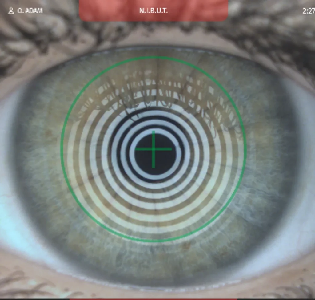
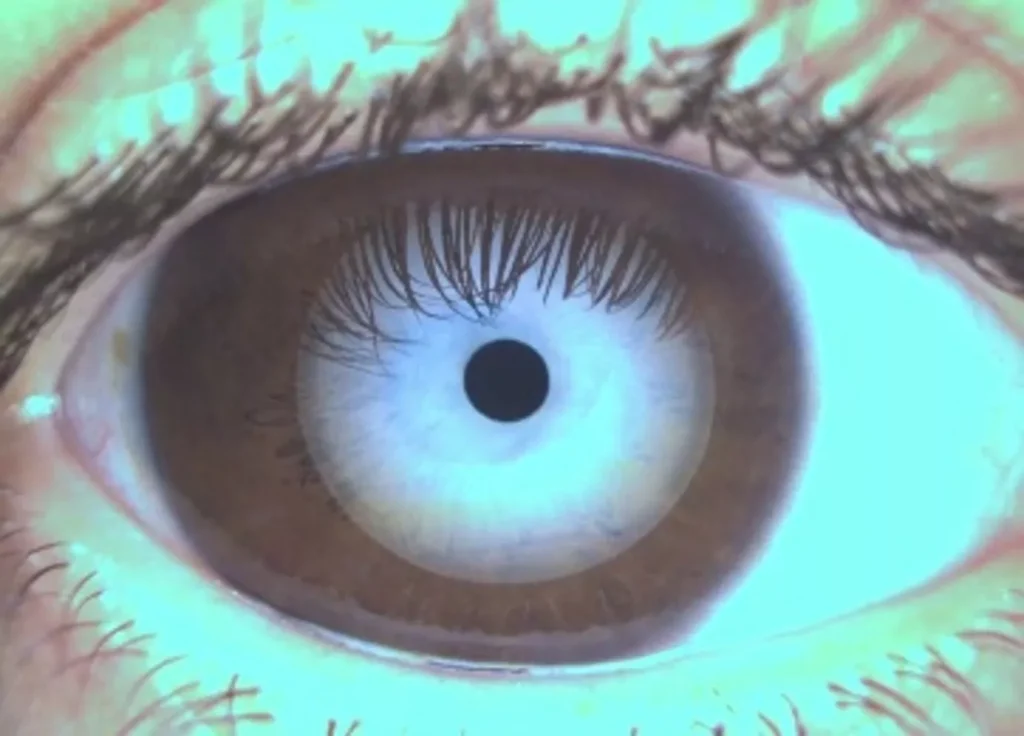
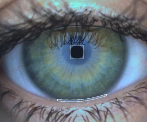
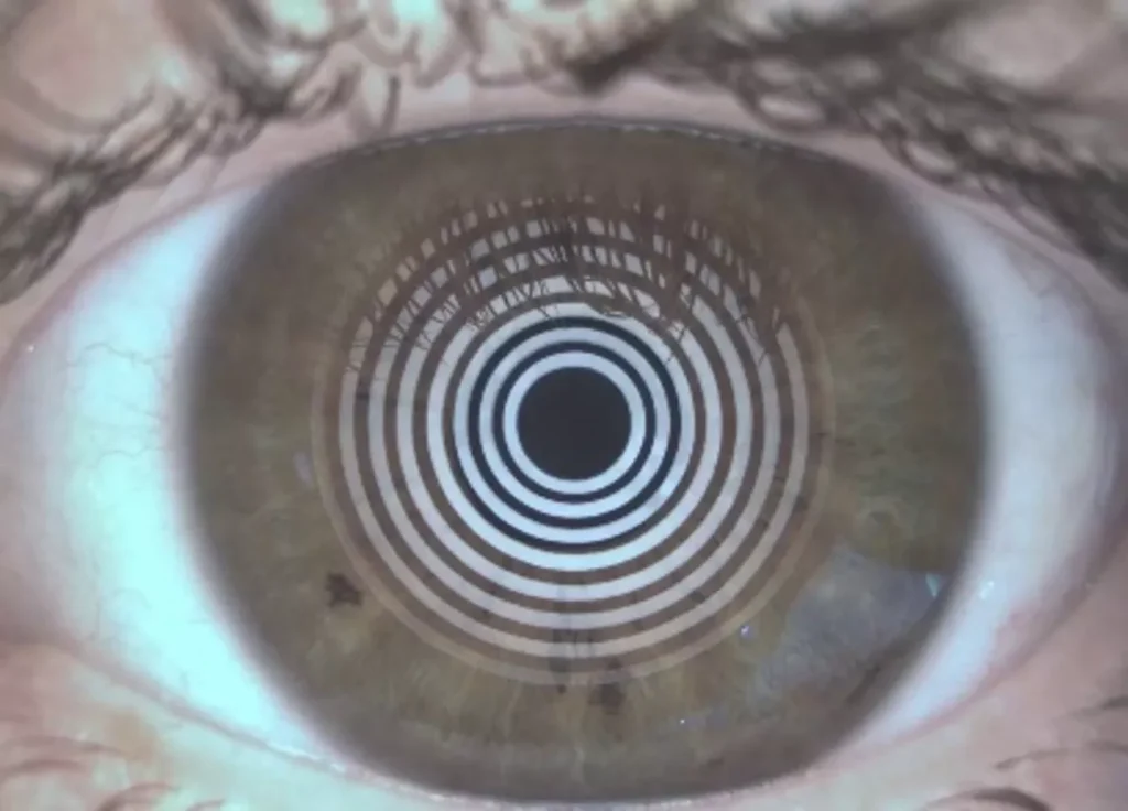
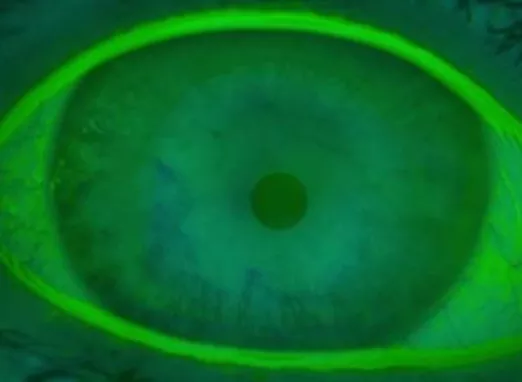
NIBUT: After a blink, this test measures how long the tear film remains on the cornea and when it breaks up, which reflects the stability of the tear film.
Lipid layer
Lipid layer: The color and structure of the lipid layer (colored fringes resembling an oil slick) allow for the assessment of the quality and thickness of the lipid layer.
Aqueous layer
Meniscus height: By measuring the height of the tear meniscus (tear at the edge of the eyelid), this test assesses the amount of water in the tears.
Mucin layer
Conjunctival staining: This test uses specific dyes (such as fluorescein) to highlight areas where mucins are deficient in order to assess the tear film’s adherence to the eye.
Meibomian glands
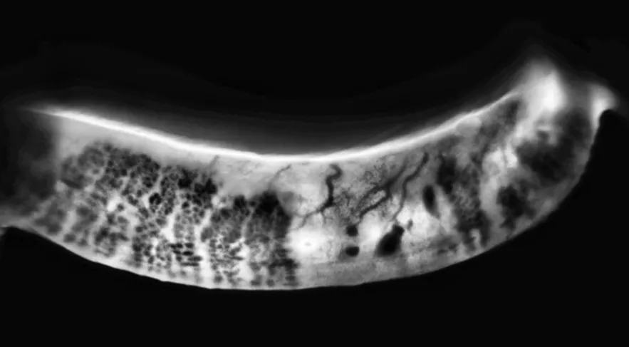
Meibography: This test provides a quick overview of the shape of the Meibomian glands. Meibography allows for the assessment of gland loss, atrophy, and obstruction, as well as telangiectasias in the eyelids.
Meibomian glands
Meibum expression: Pinching the eyelids with a special forceps to extract obstructed meibum in order to assess its quality and fluidity.
Meibomian glands
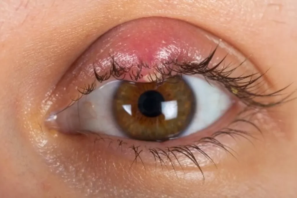
Chalazion: Observation of inflammation of a Meibomian gland
Telangiectasias of the free edge
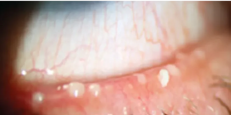
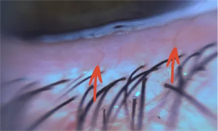
Zoom: Close-up observation of small blood vessels present on the edge of the eyelids.
Blepharitis
Zoom: Observation of eyelid inflammation
Demodex
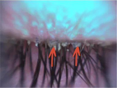
Zoom: Close-up observation of the presence of mites on the eyelid margins.
Blinking
Blink analysis: This test quantifies the number of blinks and identifies incomplete blinks.
Cellular Lesions
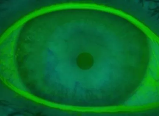
Corneal staining: This test uses specific dyes (such as fluorescein) to highlight areas where mucins are deficient in order to assess the tear film’s adherence to the eye.
Inflammation / Oxidative stress
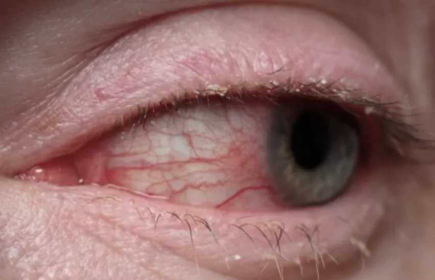
Bulbar redness: Observation of redness in the white part of the eye, a sign of irritation or inflammation of the ocular surface.
Unsure? Take the test and get your results in a few clicks
Think you may have dry eye? Take two minutes to assess your symptoms using the OSDI-6 questionnaire (six questions) provided on our site.
New IPL (intense pulsed light) treatments—all completely painless—are now available alongside a range of other treatment options for dry eye. But how much do you know about this disease?